Choosing the Right Ultrasound System

An ultrasound system is a device that uses ultrasonic waves to measure or acquire images of soft tissue or blood flow. These are mechanical waves whose frequency exceeds that of the audible frequency spectrum.

Ultrasound systems are equipped with a probe, which itself consists of a matrix of piezoelectric elements that generate an ultrasonic wave beam. These spread and are reflected on the different tissue and liquids. The wave reflects more or less depending on the organs. The main purpose of processing this wave is to measure a structure or form an image that can be used for diagnosis.

View ultrasound systems

  • How to choose an ultrasound system?

    When choosing an ultrasound system, it is very important to consider the intended applications, and of course the budget. The applications will determine certain elements.

    First, it is necessary to consider the main imaging modes that exist today:

    • B-mode ultrasound (brightness)
    • M-mode ultrasound
    • Color Doppler imaging to explore blood flow in arteries and veins
    • Ultrasound elastography to measure the stiffness of tissue

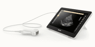
    Philips portable ultrasound system


    Then it is important to take into account the size and weight of the device:

    • Whether it is portable, on-platform or hand-held: ultrasound systems available today can easily be transported from one hospital unit to another. Many hand-held models weigh barely 500 grams and can easily be stored in a pocket or bag; some can be wireless and connected to a smartphone. They are therefore very useful for emergency and point of care medicine.
    • It is also important to take into account the size of the screen and quality of the image. Some portable ultrasound systems are able to display up to 250 shades of gray while others are equipped with color screens. Brightness is also important to consider, especially if you need to use your ultrasound system outdoors, for example in veterinary medicine. The brightness must be adjustable without compromising the readability of the observed results.
    • The type and number of transducers (shape, frequency etc.) The type of transducer you choose will depend on the intervention to be carried out. Today there are all-in-one transducers that allow a smartphone or tablet to be transformed into an ultrasound system. These are based on a downloadable application and one or more miniaturized probes (surface, abdominal, cardiac, etc.) that you connect via a simple USB port to your phone or tablet. Then you can view the results directly on your device. This type of device is suitable for emergency doctors as well as sports or humanitarian medicine.
    • Battery life is another parameter to consider, especially when choosing a portable or hand-held ultrasound system. It is best to favor a battery life of several hours for this type of device.
  • When is an ultrasound system used?

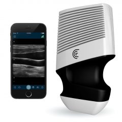
    Clarius Mobile Health portable ultrasound system

    An ultrasound system can be used to take images or measurements of different thoracic, abdominal or pelvic organs, as well as of fetuses, muscles, tendons and blood vessels. The use of endocavitary transducers allows the exploration of tissue that can’t be reached with standard probes.
    If your specialty is surgery, ultrasounds can also be used as a guide for different types of biopsy.
    On the other hand, ultrasound systems do not allow us to fully study certain organs that contain a lot of air, such as the lungs or intestines, or certain organs encapsulated in very dense structures, such as the brain. This is due to the nature of the waves used.

  • What are the main imagery modes available?

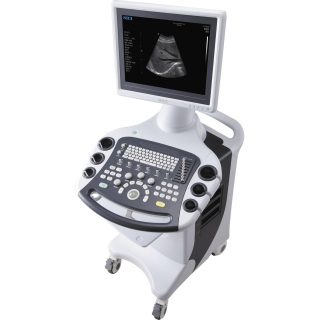
    SIUI on-platform ultrasound system

    Ultrasound modes determine to how the information will be presented to the radiologist. They define the type of image that will be displayed. The most common ultrasound modes and their applications are as follows:

    • A-mode: this is the simplest type of ultrasound. No image will be displayed on the screen, only a one-dimensional representation of reflected sound waves. This mode is used for mesuring distances.
    • B-mode (or 2B mode): linear array transducers simultaneously scan a plane through the body that can be seen as a 2D image on the screen. This is the most commonly used mode in various displines.
    • M-mode:this consists in a fast B-mode scanning sequence with successive images on the screen allowing doctors to see and measure the amplitude of movements (the perimeters of the organs producing the reflections move in relation to the transducer)
    • Doppler mode: this mode allows you to visualize and analyze blood flow, vascularization, or even the direction of flow if the Doppler is in color. This mode is used in cardiology to analyze the condition of heart valves or the vascularization of certain tissue.
    • Elastography mode: with this mode it is possible to measure the hardness of tissue and to diagnose, for example, a potential hepatic fibrosis.
    • 3D and real-time 3D (4D) mode: this mode is used during ultrasound examinations of pregnant women to obtain a three-dimensional image of the fetus and observe its movements in utero. But the risks to the fetus are not negligible (see question 2).
  • Why is an ultrasound gel used?

    In order to obtain detailed images from an ultrasound system, the ultrasound used must be very high-pitched, i.e. at very high frequencies between 3 and 20 MHz. However, at such frequencies, ultrasound is no longer transmitted through the air. Therefore, a substance capable of expelling air between the transducer and the patient’s skin must be used. This is why, as we saw in question 3, an ultrasound system can’t help in the study of organs that contain a lot of air.

    Using an ultrasound gel allows ultrasound to be transmitted without modifying it. Pure water could also be used in the same way, but a gel is more practical because it doesn’t leak, doesn’t get the patient wet, serves as a lubricant and seals the roughness between the transducer and the skin. During certain procedures, a disinfectant such as povidone-iodine can be used instead.

    Ultrasound gel, which is applied generously to the belly of a pregnant woman for a prenatal ultrasound, for example, is composed of purified water, a gelling agent and preservatives (antibacterial). In general, it is neither greasy nor tinted and does not leave any stains or marks on the patient’s clothing. It is not advisable to warm it up because a warmer temperature will reduce its cohesiveness and promote the development of germs.

RELATED GUIDES
1 Star2 Stars3 Stars4 Stars5 Stars (No Ratings Yet)
Loading...
1 comment
  • Kristofer Van Wagner says:

    I totally agree that medical practitioners should consider the size of the screen for an ultrasound machine as well as taking into consideration the quality of image that it produces because it is imperative for many diagnostics. I remember when I took my wife for her antenatal check-up and we were very pleased to know that the maternity hospital is equipped with an ultrasound machine that can display up to 250 shades of grey. I do believe that medical practitioners should take this into consideration when buying and ultrasound machine.

Leave a Reply

Your email address will not be published. Required fields are marked *