A flat panel detector is made up of a semiconductor matrix and allows a digitized image to be aquired when taking an X-ray. It therefore enables the replacement of traditional X-ray film in order to simplify the process.


A flat panel detector is made up of a semiconductor matrix and allows a digitized image to be aquired when taking an X-ray. It therefore enables the replacement of traditional X-ray film in order to simplify the process.
The flat panel detector has advantages over film such as sensitivity and better process reproducibility, but it also has disadvantages such as cost and lower spatial resolution. Here is a more comprehensive list of the advantages and disadvantages:

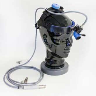
Canon portable flat panel detector
In order to make the best possible choice when purchasing a flat panel detector, several criteria such as image quality, application and the size of the device must be taken into account. Here is the list of criteria:
It is difficult to determine exactly how the performance of flat panel detectors will change over time, even though some studies have been carried out on their ageing process.
It should simply be noted that the sensitivity of scintillator detectors decreases over time and depends primarily on the number of images taken
For SwissRay detectors, for example, the service life is at least five years. Canon detectors are guaranteed for 210,000 shots and seven years of service life.
The principle of a flat panel detector is to transform an X-ray beam of energy ranging from 20 to 120 keV into an electrical signal, which is then digitized. There are three main operating techniques for direct conversion and indirect conversion flat panel detectors. These three technologies depend on the number of steps required to transform an X-ray photon beam into an electrical signal.
Please note: we have not presented the following in this buying guide:
Each of the three techniques discussed in the previous section has its advantages and disadvantages. They are outlined below:


 (4 votes, average: 2.50 out of 5)