A rigid endoscope is often used for applications where access to the inspection area is not very sinuous, such as cranial sinuses, the ear canal, urinary devices, etc. It is coupled to a light source and sometimes a video camera is attached to the lens.
It can have a wide-angle lens, which allows for wide framing of close objects. It also offers high-definition images thanks to transmission by optical lenses. The rigid structure of the endoscope is due to the succession of lenses aligned in the tube. This allows the practitioner to obtain a clear image of the inspected area.
Lastly, rigid endoscopes can have one or more channels through which an endoscopic instrument can pass. These can be forceps to grasp or remove foreign bodies or tissue samples; scissors to cut tissue; brushes to collect cells or a lace to catch polyps for example.
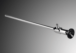
Hipp Endoskop Service laparoscope