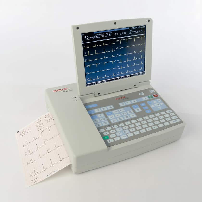
A Schiller 12-channel electrocardiograph
The number of electrodes is decisive for the accuracy of the diagnosis: the higher the number, the more reliable and accurate it will be.
An electrocardiograph with four leads (three electrodes and one ground) allows for basic monitoring. It can measure the heart rate, analyze the change in heart rate, visualize P waves, QRS complex and T waves. Usually, electrodes are placed on the wrists and ankles. The electrodes on both arms and the left leg reflect the electrical variations of the heart while the electrode on the right leg serves a ground.
An electrocardiograph with 12 leads provides a more precise and accurate diagnosis by eliminating noise and disturbance from some leads. It also makes it possible to refine the diagnosis on a specific part of the heart. A standard 12-lead electrocardiograph includes six peripheral leads (also called limb leads) and six precordial leads (also called chest leads).