Choosing the Right Anatomical Model

An anatomical model is a real and accurate representation of an anatomical structure. It can be made from a variety of materials to be as realistic as possible and thus make it easier to illustrate a medical procedure, for example. This guide only deals with human anatomical models and does not cover veterinary anatomical models.

View our anatomical models

  • What are anatomical models used for?

    Anatomical models are used for three main purposes: preoperative planning, training, and communication.

    • Pre-operative planning: Anatomical models are a valuable resource for planning complex surgical procedures or for checking whether or not the shape and size of an implant are suitable before placement. There are anatomical models for heart surgery, vascular surgery, and implantology, just to name a few.
    • Training: Anatomical models are also used in teaching and learning activities, particularly for training health professionals in schools, universities, and training centers. Training anatomical models are excellent teaching tools for studying anatomy and physiology in detail. They are an effective and convenient alternative to using cadavers to practice and develop medical and nursing skills.
    • Communication: Anatomical models are also excellent communication tools, particularly for demonstrating new medical devices and training professionals in their use.
  • What materials are anatomical models made of?

    Anatomical models can be made of plastic, organic materials such as wood, or foam. There are also virtual anatomical models and 3D-printed models.

    • Silicone anatomical models: Plastic anatomical models are the most common, especially silicone ones, as they don’t require special equipment and are easy to mold. However, silicone models cannot be used in certain situations, particularly for demonstrating procedures that use ultrasounds. Since silicone attenuates ultrasound, objects appear to be located deeper than they actually are.
    • Anatomical models made from organic materials and synthetic materials (other than silicone): These can be used for ultrasound procedures and are therefore a good alternative to silicone models.
      • Anatomical models made from organic materials, such as gelatine and agar-agar gel, are potentially cheaper options, as they have lower production costs, however, they tend to dry out and deteriorate over time. There are also wooden anatomical models, but these are mainly used for decorative purposes.
      • Anatomical models made of synthetic materials, such as polyvinyl chloride (PVC) and polyvinyl alcohol (PVA), last longer than models made of organic materials. In addition, they can be used several times for minimally invasive procedures, such as needle insertion. However, they usually require special equipment to be manufactured.
    • Foam anatomical models: Some anatomical models are made of foam to reproduce the sponginess of certain tissues.
    • Virtual and 3D-printed anatomical models: Digital (virtual) anatomical models are designed and viewed with specific software. 3D design and printing have come a long way and there are many 3D-printed anatomical models, particularly in universities. These are practical and easy to use, but also very realistic, as they are based on 3D digital images taken from real human bodies. Their main drawback is their high cost.
  • What parts of the body are represented in anatomical models?

    Anatomical models can represent practically any part of the human body. There are whole-body models and models of specific parts, which cover the different organ systems (skeletal, muscular, cardiovascular, digestive, nervous, respiratory, etc.) and their constituents. They can be used to teach and train a wide variety of medical procedures.

    • Skeletal system: Skeleton models can display the bone structure of the entire body or just a part of the body, as well as tendons, ligaments, and other elements of the skeletal system.
    • Muscular system: Muscle models show skeletal, cardiac and/or smooth muscles, the latter forming the outer layer of some organs.
    • Cardiovascular system: Heart models are very useful for teaching/learning and for planning cardiology surgeries.
    • Digestive system: Digestive system models include the oral cavity, esophagus, stomach, intestines, liver, gallbladder, and pancreas. Stomach and intestine models are widely used in gastroenterology.
    • Urinary system: Urinary system models display the kidneys, urethra, bladder, ureters, and other organs and glands connected to the urinary system.
    • Reproductive system: Anatomical models of the male and female reproductive systems can include the genital organs and glands responsible for human reproduction.

  • What other characteristics should you consider when choosing an anatomical model?

    Depending on the intended use, other characteristics may be important when choosing an anatomical model. These include its level of detail, whether or not it can be taken apart, transparency, and how easy it is to clean.

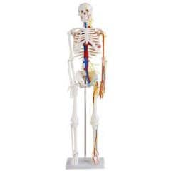
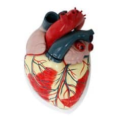
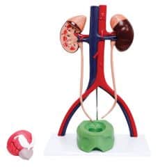
    • Model size and level of detail: Choosing an anatomical model with the right level of detail for the intended use is essential. For example, a two-part brain model will allow you to see each of the hemispheres but not the lobes, while a nine-part model will provide a fully detailed view, although it may be too complex for certain levels of education. In all cases, the various areas must be easy to identify in the model. To help distinguish between them, different colors are generally used. Some models display both the organs and the blood vessels.
    • Models that can be partially or completely disassembled: Certain anatomical models can be partially or completely taken apart, allowing the outside and inside of the anatomical structure to be seen. This is particularly useful for lessons in which students must inspect a particular organ.
    • Transparent models: If you can’t use a model that can be taken apart, you can use transparent anatomical models, which also allow you to see inside the organs.
    • Easy-to-clean anatomical models: Cleaning anatomical models regularly is essential to keep them in good condition and reduce the risk of contamination. Simply wipe them with a soft cloth dampened with detergent. It is not advisable to use corrosive products, as these can cause the model’s colors to fade or erase any inscriptions.
RELATED GUIDES
1 Star2 Stars3 Stars4 Stars5 Stars (No Ratings Yet)
Loading...
No comment

Leave a Reply

Your email address will not be published. Required fields are marked *