Choosing the Right Catheter

A catheter is a long, thin tube with a diameter adapted to the part of the body it is used for. Inserted into a vessel or body cavity, they are mainly used to dispense fluids, such as medical solutions, into the body and to insert or remove medical devices such as stents or pacemaker electrodes. Some catheters are also used to measure physiological parameters such as blood pressure.

View catheters

  • What are the different parts of a catheter?

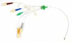
    A catheter is a tube with one end made up of one or more ports for connecting tubing or inserting an instrument.

    There are catheters made up of several internal channels or “lumens.” They allow different fluids to be administered simultaneously via a single catheter.

    Some catheters also have a balloon. Once inflated, the balloon holds the catheter in place in a cavity. This is the case, for example, for the Foley catheter, which is held in place by an inflated balloon in the bladder neck. To collect urine, this type of catheter can be connected to a urine bag.

  • What are the different types of catheters?

    Vygon access catheter

    A wide variety of catheters exist according to their use. The three main types of catheters are:

    • Infusion catheters (venous or arterial): allow one or more fluids such as medical solutions to be administered in the body.In this case, the catheter is placed in a vein or artery for ongoing administration to avoid having to constantly prick the patient. Infusions of medical solutions can be prolonged or repeated. For example, during childbirth, a pregnant woman may be placed on an infusion to receive a local anesthetic that will reduce labor pain. The anesthesiologist places the catheter in the epidural space at the bottom of the spine and injects the solution containing the local anesthetic through this tube.
    • Urine drainage catheters: drain urine from the bladder or kidneys of patients with bladder weakness or who are unable to urinate. This may be due to a calculus, a tumor or the after-effects of surgical treatment. These catheters can be permanent or intermittent. Some are inserted through the urethra, such as a Foley catheter, or directly into the kidney through a percutaneous method, such as a nephrostomy.
    • Angioplasty catheters: These catheters allow a vessel to be dilated with the balloon attached to the tip. They are mainly used to treat occlusions or arterial stenoses (coronary arteries, peripheral arteries, etc.).
  • What is a hydrophilic catheter?

    The term “hydrophilic” is used to refer to one of the fundamental characteristics of the outer coating of the catheter, which is generally smooth, uniform and immersed in a saline solution.

    The main purpose of a hydrophilic catheter is to limit the sensation of pain, pressure or discomfort that a patient might feel when the tube is inserted. As they are pre-lubricated and optimally hydrated, they decrease the risk of urethral lesions by reducing friction.

    In the context of intermittent aseptic catheterization, available studies show that in practice the use of hydrophilic catheters appears to be preferable. In particular, compared to standard catheters, they reduce bacteriuria (presence of bacteria in the urine) as well as long-term urethral complications, such as urethral stenosis.

  • What are the risks associated with using a venous catheter?

    The peripheral venous catheter is usually used in emergency situations, such as resuscitation, or more commonly to administer intravenous treatments.

    Its installation is often a vital necessity, but it is not a procedure without consequences and it involves certain risks. 15% of patients who undergo this procedure develop a complication.

    The main risks associated with using a venous catheter are:

    • Infection (nosocomial diseases): catheter infections are estimated to represent 18 to 25% of nosocomial infections. Additionally, nearly 80% of contracted nosocomial diseases are reported to occur in catheterized patients. The incidence of infection appears to be higher for the central venous pathways than for the peripheral venous pathways.
    • Pneumothorax: this can occur immediately or within 48 hours of catheter insertion. The pleural cavity, between the lungs and the rib cage, fills with air, causing respiratory discomfort, coughing, chest pain and even feelings of anxiety. This can be resolved within a few weeks or, when necessary, it can require hospitalization to drain the air from the chest.
    • Hemorrhages by venous vascular injury: these generally occur on the internal jugular and femoral tract and remain mild. Ultrasound guiding reduces this risk.
    • Thrombosis: this complication, less well known than catheter infection, is often a silent disease so it is more complicated to diagnose. Nevertheless, thrombosis and infection are often linked. The detection of a thrombus doubles the risk of catheter-related infections.
  • How is a catheter inserted?

    Catheter insertion is in most cases performed without analgesia. Some catheters are inserted directly into the cavity, such as the urethral catheter, others are inserted percutaneously as seen in the case of a nephrostomy. The length and diameter of the catheter depend on the vessel or cavity to be reached, as well as the age and sex of the patient.

    The pain experienced during insertion varies from patient to patient. It depends on the application, the type of catheter used (hydrophilic or not) and the physical state of the patient (age, weight, physical condition and pain resistance). It also depends on the health professional who performs the procedure as it requires a certain amount of experience and dexterity.

RELATED GUIDES
1 Star2 Stars3 Stars4 Stars5 Stars (No Ratings Yet)
Loading...
No comment

Leave a Reply

Your email address will not be published. Required fields are marked *